Interview with Dr. Buelow: More than meets the eye
Blog post
September 8, 2020
At his second visit to an emergency veterinary center, Fabio, a young pug, was fortunate to have been observed by Dr. Mary Buelow of Animal Dentistry & Oral Surgery of Leesburg, Virginia. Dr. Buelow, a board-certified veterinary dentist, saw symptoms that led her to believe that the case was more than it seemed on the surface. Our interview with Dr. Buelow explains how the more detailed dental anatomy taken on a Planmed Verity® CBCT can lead to a precise diagnosis.
Seeing more
Fabio visited the adjoining veterinary emergency clinic two days prior with extreme swelling and discharge from his eye. The treatment of antibiotics did not result in any improvement, and the pug returned to the ER two days later for further treatment.
Dr. Mary Buelow happened to see the pug in the emergency room, and from previous experience seeing ocular discharge and swelling caused by dental disease, Dr. Buelow suggested dental imaging. She knew that images taken with the Planmed Verity® would give the most detailed anatomical view to determine the correct course of action.
Although pustular ocular discharge and conjunctival swelling are rare presentations of dental disease, Dr. Buelow knew that the volumetric images would be the optimal path for an accurate analysis.
Fabio with extreme swelling and discharge from his eye.
The root of the problem
Dr. Buelow began with a standard 2D X-ray image using a phosphor plate system, as is commonly the only dental imaging performed in private and specialty practices. However, no abnormalities were noted on the dental X-rays that would explain the extreme swelling.
Intraoral images showed no abnormalities.
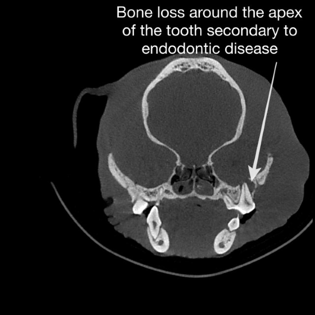
3D images captured with the Planmed Verity CBCT confirmed the diagnosis of severe endodontic disease, causing a tooth root “abscess.” Rapid capture of a CBCT image had the clear advantage of uncovering the pathology that caused the facial swelling over the intraoral X-ray, which appeared normal.

Without the rapid 3D image capture, what would have been the next course for Fabio? Traditional CT scans can be an option, but ultimately, the large slices of a CT scan image are not ideal for diagnosing dental disease compared to the thin, sharp image slices of CBCT image. The CT imaging process involves more time, delaying diagnosis and treatment, and more costly for the pet parent.
Dr. Buelow explained, "This is a serious issue for brachycephalic breeds. We should not be treating these breeds without CBCT as traditional dental x-rays miss too much pathology. Fabio is an excellent case to justify that CBCT is a more sensitive diagnostic tool, not only for brachycephalic breeds but for all of our patients."
From a scan complete in 30 seconds, Dr. Buelow detected a tooth abscess and several other dental issues. The ADOS staff immediately prepared Fabio for his surgery to be performed by Dr. Matthew Raleigh DVM.
Fabio four days after the dental surgery in which the diseased tooth was removed.
Proud of "Pinky"
Animal Dentistry & Oral Surgery is proud to present the Planmeca Verity® CBCT that they have named "Pinky" as a member of the team. A dedicated “Pinky” page on the clinic's website offers extensive information CBCT technology’s benefits. Advancements in medicine have prompted pet parents to expect and embrace the same innovations in veterinary medicine – and veterinary dentistry – for their pets.
Dr. Buelow concurs. "As dental specialists, we are in a fortunate position when veterinarians and their clients come to us for help. We have chosen not to price our CBCT scans significantly more than an X-ray to make it an affordable option for our clients. Often, we include a CBCT as a required part of a treatment plan. Our clients are pleased with the results that CBCT technology brings to plan a course of treatment to ease their pet's pain and regain their health."
Animal Dentistry & Oral Surgery, Inc is a veterinary dental specialty practice established in 1999 to serve the oral surgical and dental health needs of companion animals in the northern Piedmont area of Virginia.
Dr. Mary E. Buelow DVM, DAVDC practices at the Animal Dentistry & Oral Surgery in Leesburg, Virginia. A veterinarian for over 15 years, Dr. Buelow completed her residency in veterinary dental and oral surgery at the University of Illinois and has earned her board certification from the American Veterinary Dental College. Dr. Buelow has practiced in New York and served as an active member and President of the Veterinary Medical Associate of New York City. After serving as an Assistant Professor of Clinical medicine and practicing clinician at the University of Pennsylvania School of Veterinary Medicine, Dr. Buelow relocated to the Virginia as a dental specialist at Animal Dentistry & Oral Surgery. Dr. Buelow has not been compensated by the Planmeca Group for her participation in this content.


